Are you looking for an answer to the topic “Does lumbar MRI include sacroiliac joint?“? We answer all your questions at the website Musicbykatie.com in category: Digital Marketing Blogs You Need To Bookmark. You will find the answer right below.
Magnetic resonance imaging (MRI) can reliably detect inflammation and structural changes in sacroiliac joints (SIJs) in patients with lower back pain (LBP). However, patients with LBP are usually referred for MRI of the lower back (e.g. lumbar spine LS), and imaging of the SIJs is rarely requested for these patients.A lumbar MRI specifically examines the lumbar section of your spine — the region where back problems commonly originate. The lumbosacral spine is made up of the five lumbar vertebral bones (L1 thru L5), the sacrum (the bony “shield” at the bottom of your spine), and the coccyx (tailbone).MRI is the most sensitive imaging technique to detect sacroiliitis. It is the only imaging modality that can reliably reveal bone marrow oedema and inflammation around the sacroiliac joints and is comparable to low dose CT for demonstrating erosions and ankyloses (13).

Table of Contents
Does a lumbar MRI include the sacrum?
A lumbar MRI specifically examines the lumbar section of your spine — the region where back problems commonly originate. The lumbosacral spine is made up of the five lumbar vertebral bones (L1 thru L5), the sacrum (the bony “shield” at the bottom of your spine), and the coccyx (tailbone).
Can MRI show sacroiliac joint?
MRI is the most sensitive imaging technique to detect sacroiliitis. It is the only imaging modality that can reliably reveal bone marrow oedema and inflammation around the sacroiliac joints and is comparable to low dose CT for demonstrating erosions and ankyloses (13).
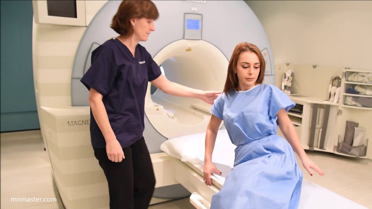
Does MRI Show an SI Joint Problem?
Images related to the topicDoes MRI Show an SI Joint Problem?
Is the sacroiliac joint part of the lumbar spine?
The sacroiliac joint connects the hip bones (iliac crests) to the sacrum, the triangular bone between the lumbar spine and the tailbone (coccyx). The primary function of the sacroiliac joints is to absorb shock between the upper body and the pelvis and legs. The sacroiliac joint typically has little motion.
What shows up on a lumbar spine MRI?
A lumbar spine MRI can detect a variety of conditions in the lower back, including problems with the bones (vertebrae), soft tissues (such as the spinal cord), nerves, and disks.
Will a lumbar MRI show hip problems?
Occasionally, hip pathologies may present alone or combined with lumbar spine pathology, especially lumbar stenosis. Although the history and clinical examination may help differentiate between the two, hip X-rays alone without accompanying magnetic resonance imaging (MRI) studies may prove unreliable.
Does lumbar spine MRI show coccyx?
A lumbar spine MRI focuses on the L1 – L5 vertebrae of the lumbar spine and the surrounding nerves, soft tissues and blood vessels. It is possible to see portions of the coccyx in a lumbar scan however it is not the main focus. It is possible to have a lumbosacral MRI scan to include the coccyx.
What kind of MRI is used for sacroiliitis?
In cases of suspected infective sacroiliitis, our imaging protocol is more extensive than that for suspected inflammatory sacroiliitis and, in this setting, we will routinely perform oblique coronal (T1SE and T2 fat-sat) and oblique axial sequences (T1SE, T2 fat-sat and T1-fat-sat post contrast) images.
See some more details on the topic Does lumbar MRI include sacroiliac joint? here:
Should a Lumbar MRI for back pain routinely include the sacro …
Conclusions: We conclude that routinely imaging the SIJ in MRI lumbar spine series is not cost-effective or a useful use of resources. The SIJ should be imaged …
Diagnosis for Sacroiliac Joint Pain – WebMD
X-rays can help them look for changes in the SI joint. Computed tomography (CT), a powerful X-ray scan, can give them even more details.
What Does a Lumbar Spine MRI Show? – American Health …
Some of the inconsistencies that a lumbar spine MRI may show include compression or inflammation of the spinal cord and adjacent nerves, …
Imaging the Patient With Sacroiliac Pain – ScienceDirect
The SI joints have a unique anatomic layout and composition and can be imaged with a variety of techniques including conventional radiographs, computed …
How do you test for sacroiliac joint?
The surest way for a doctor to know if you have SI joint dysfunction is through an injection of numbing medicine into your joint. An X-ray or ultrasound guides the doctor to where to put the needle in. If the pain goes away after the shot, you know the joint is the problem.
Can sacroiliitis be missed on MRI?
Conventional radiography is frequently normal in early sacroiliitis. Similarly, bone scintigraphy has low sensitivity, and MRI can miss up to one-third of the patients with AxSpA as well.
How do I know if my back pain is sacroiliac?
- Pain on one side of your lower back.
- Burning sensations or stiffness in your pelvis.
- Pain that doesn’t extend above your waistline.
- Pain that radiates into your hip, groin, or thighs.
- Pain that is worse when you stand or walk.
Is L5 S1 the same as SI joint?
The L5 and S1 nerves are near the SI joint and studies have shown that SI joint dysfunction can cause pain and other symptoms in the distribution of these nerves. The SI joint is separate from the sciatic or spinal nerve(s); however, the SI joint can cause sciatica-like symptoms.
What does sacroiliac joint pain feel like?
You may experience sacroiliac (SI) joint pain as a sharp, stabbing pain that radiates from your hips and pelvis up to the lower back and down to the thighs. Sometimes it may feel numb or tingly, or as if your legs are about to buckle.
MRI lesions in the sacroiliac joints of patients with spondyloarthritis
Images related to the topicMRI lesions in the sacroiliac joints of patients with spondyloarthritis

Does lumbar spine MRI show piriformis?
MRI can help to correctly diagnose pirifor- mis syndrome and also to differentiate piri- formis syndrome from other possible causes of lower lumbar pain and sciatica, such as lumbar disk herniation, lumbar stenosis, and mass lesions in the region of the piriformis muscle [5].
What will an MRI show for hip pain?
An MRI can reveal fraying or tears of the cartilage and labrum. Sometimes it is necessary to find a way to differentiate pain radiating from the hip joint and pain radiating from the lower abdomen. To accomplish this, the hip can be injected with a steroid analgesic.
Does MRI show inflammation in back?
Conclusions. Inflammatory MRI lesions in early AS are seen more often in posterior structures of the spine. This may be relevant for the diagnosis of early AS and the early detection of inflammatory spinal involvement.
Does hip MRI show SI joint?
An MRI of your pelvis/hips can be useful in investigating pain, weakness or swelling of joints. An MRI can do this by examining bones, joints, and soft tissues such as cartilage, muscles, and tendons for injuries or the presence of structural abnormalities. We can scan your hips, pelvis and Sacroiliac (SI) joints.
Does pelvic MRI show hip joint?
Pelvic MRIs allow a doctor to see the area between your hips, your reproductive organs, blood vessels, and hips themselves.
Does lumbar spondylosis cause hip pain?
The vertebra may slip out of place on the vertebra positioned below placing pressure on nerves that may cause leg pain or lower back pain. Because spinal stenosis with spondylolisthesis can cause significant hip or knee pain, the presence of degenerative arthritis at these joints must be considered.
Will pelvic MRI show sacrum?
Although sacrum is not routinely covered completely on L-spine MRI, our study revealed that L-spine MRI was the most common modality that detected SIF (n = 27, 64.3%), followed by pelvic bone CT (n = 6, 14.3%), bone scan (n = 5, 11.9%), and pelvis MRI (n = 4, 9.5%).
What does an MRI of the sacrum and coccyx show?
MRI of the sacrum/coccyx is also an effective tool to evaluate for disorders of the sacroiliac (SI) joints, the joints that connect the lower spine to the pelvis. Both inflammatory and degenerative conditions can affect the SI joints and cause pain in the region.
What kind of MRI is used for tailbone pain?
Lumbosacral MRI would be most helpful in cases in which the coccygeal pain is suspected to be referred from anatomic structures located more superiorly within the spine (ie, at the lumbar or sacral regions).
Which of the following tests checks for sacroiliac joint dysfunction?
- Gaenslen Test.
- FABER / Patrick Test.
- Thigh Thrust / Femoral Shear Test.
- ASIS Distraction.
- Sacral Compression.
Lumbar spine MRI scan, protocols, positioning and planning
Images related to the topicLumbar spine MRI scan, protocols, positioning and planning
What kind of doctor do I see for sacroiliac joint pain?
A clinician such as a physical therapist, pelvic health specialist, or pain management specialist can perform these tests to help you diagnose SI joint disease or SI joint dysfunction.
How can I tell if I have sacroiliitis?
Common symptoms of sacroiliitis include pain that: Occurs in the lower back, buttock, hip, or thigh. Gets worse after long periods of sitting or standing, or getting out of a chair. Worsens after rotating your hips.
Related searches to Does lumbar MRI include sacroiliac joint?
- 3 tests to tell you if your back pain is caused by si
- what will mri of si joint show
- does mri of lumbar spine show sacroiliac joint
- does lumbar spine mri show pelvis
- mri sacroiliac joint
- how to read mri of si joint
- does lumbar mri include sacroiliac joint
- does a lumbar mri include the si joint
- will lumbar mri show si joints
- si joint dysfunction self test
- does a lumbar mri show si joints
- mri sacroiliac joint ankylosing spondylitis
- does mri lumbar spine include si joints
- si joint mri protocol
- does lumbar spine mri show hips
- does lumbar mri show sacroiliac joint
Information related to the topic Does lumbar MRI include sacroiliac joint?
Here are the search results of the thread Does lumbar MRI include sacroiliac joint? from Bing. You can read more if you want.
You have just come across an article on the topic Does lumbar MRI include sacroiliac joint?. If you found this article useful, please share it. Thank you very much.